Anatomia 2024, 3(2), 124-135; https://doi.org/10.3390/anatomia3020010 - 3 Jun 2024
Abstract
►
Show Figures
The pancreas is a vital organ nestled deep within the abdomen, playing a crucial role in both endocrine and exocrine functions. It is elongated and tadpole-shaped, with a head, body, and tail. The intricate connections to adjacent structures through a network of blood
[...] Read more.
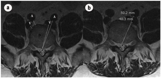
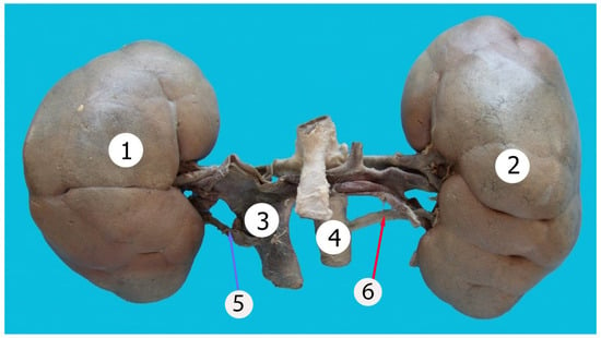
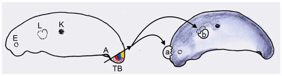
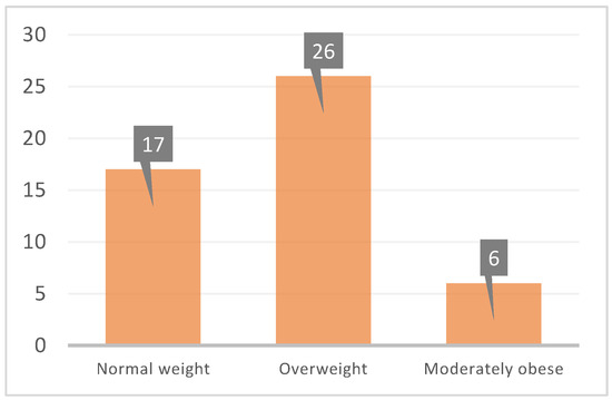
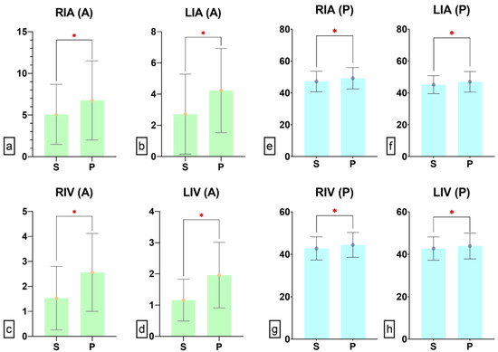
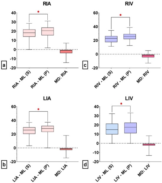
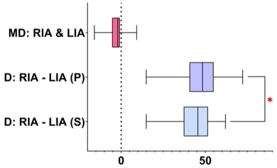
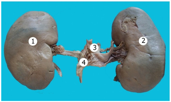
The pancreas is a vital organ nestled deep within the abdomen, playing a crucial role in both endocrine and exocrine functions. It is elongated and tadpole-shaped, with a head, body, and tail. The intricate connections to adjacent structures through a network of blood vessels, ducts, and supportive tissue transform pancreatic cancer into one of the most fatal malignancies globally as a result of a typically late diagnosis and metastatic form of the disease. Lymph node metastasis (LNM) is prevalent in the majority of individuals diagnosed with pancreatic cancer, signifying a critical factor influencing prognostic outcomes. The para-aortic lymph nodes (PALN) play an important role in the lymphatic drainage of various organs, including the kidneys, pancreas, and parts of the gastrointestinal tract. In pancreatic cancer, the risk of PALN metastasis holds considerable clinical significance, and diagnosing your involvement is primordial to therapeutic decisions and to increase the survival expectations of these patients.
Full article

Figure 1


